Anatomy Pictures Of Lower Back And Hip : Sacroiliac (SI) Joint Dysfunction | Houston Methodist. In vertebrate anatomy, hip (or coxa in medical terminology) refers to either an anatomical region or a joint. This mri hip joint axial cross sectional anatomy tool is absolutely free to use. Understanding lower back anatomy is key to understanding the root of lower back and hip pain. Tired of tight, stiff lower back and hip muscles? Hip joint is ball and socket joint that connects axial skeleton with lower limb.
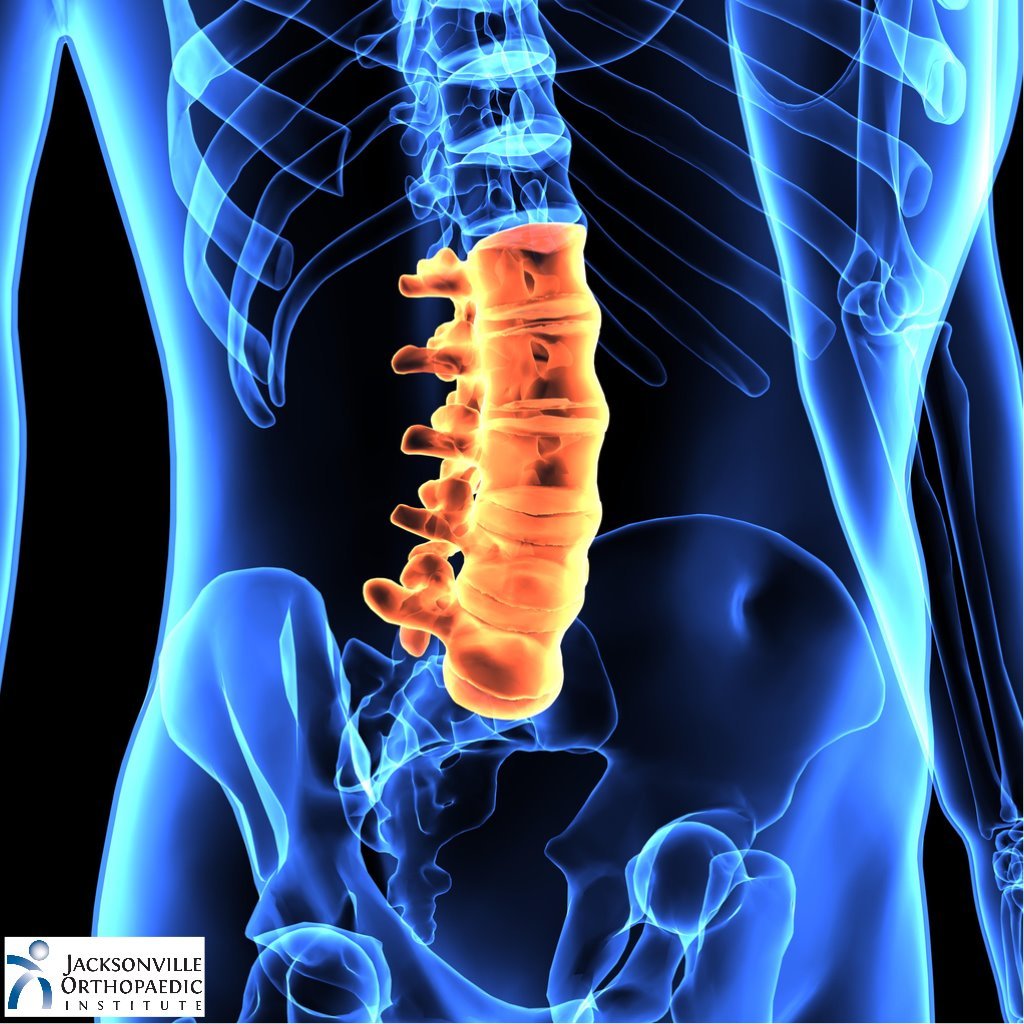
A collection of anatomy notes covering the key anatomy concepts that medical students need to learn. Muscles of the lower limb | anatomy model. Anatomy pictures of lower back and hip / … The human spine is composed of 4 sections of vertebrae. Use the mouse scroll wheel to move the images up and down alternatively use the tiny arrows (>>) on both side of the image to move the images.
Suffering Back Pain? How is your Sacroiliac Joint function? — JUICE BOSS HQ from static1.squarespace.com A collection of anatomy notes covering the key anatomy concepts that medical students need to learn. Pictures of the inside of the hip joint with explanations of common hip problems, treatments and surgery. While the thigh muscles will be slip into the anterior, medial and posterior groups. Discover the relationship between hip dysplasia and hip anatomy and see hip anatomy pictures. Knee assessment and hip mechanics online course: Possible causes of lower back and hip pain include sprains, strains, and a herniated disk. Ankle, aorta, back, backwards, bite, blue+eyes, body, body parts, bone, brain, brown eyes, butt, calf, calves, cartoon eyes. Right superficial inguinal lymph nodes.
As long as you're not injured, doing strengthening exercises, plus dr.
Bursae of the lower limb: Interactive and dynamic anatomical atlas of anatomy. These sections are cervical (neck), thoracic (upper and middle back), lumbar (lower back), and sacrum (tailbone). A collection of anatomy notes covering the key anatomy concepts that medical students need to learn. Ankle, aorta, back, backwards, bite, blue+eyes, body, body parts, bone, brain, brown eyes, butt, calf, calves, cartoon eyes. Understanding lower back anatomy is key to understanding the root of lower back and hip pain. The anatomy of the fascia lata and iliotibial tract. Learn about anatomy lower limb with free interactive flashcards. Knee assessment and hip mechanics online course: Pictures of the inside of the hip joint with explanations of common hip problems, treatments and surgery. Want to learn more about it? The hip muscles are going to be slip into hip muscles and gluteal muscles. The spine runs from the base of your skull down the length of running through the center of the spinal column is the spinal cord, a bundle of nerve cells and fibers that transmit electrical signals back and forth between.
Anatomy pictures of lower back and hip / … The socket is a concave depression in the lower side of the pelvis (also called the acetabulum). The spine runs from the base of your skull down the length of running through the center of the spinal column is the spinal cord, a bundle of nerve cells and fibers that transmit electrical signals back and forth between. In vertebrate anatomy, hip (or coxa in medical terminology) refers to either an anatomical region or a joint. The iliopsoas muscle, which extends from the lower back to.
Lower Back Muscle Anatomy and Low Back Pain from ix-cdn.brightedge.com And you'll be in a better position to help your doctor pinpoint the cause. She is a former american college of sports medicine certified personal trainer and currently works as a level 1 crossfit coach. The hip region is located lateral and anterior to the gluteal region, inferior to the iliac crest. Anatomy pictures of lower back and hip / … Bursae of the lower limb: Muscles of the lower limb | anatomy model. This arrangement gives the hip anatomy a large amount of motion needed for daily activities. These sections are cervical (neck), thoracic (upper and middle back), lumbar (lower back), and sacrum (tailbone).
Possible causes of lower back and hip pain include sprains, strains, and a herniated disk.
Related online courses on physioplus. By dr arun pal singh. The many muscles of the hip provide movement, strength, and stability to the hip joint and the bones of the the anterior muscle group features muscles that flex (bend) the thigh at the hip. While the thigh muscles will be slip into the anterior, medial and posterior groups. Continue scrolling to read more below. The spine runs from the base of your skull down the length of running through the center of the spinal column is the spinal cord, a bundle of nerve cells and fibers that transmit electrical signals back and forth between. And you'll be in a better position to help your doctor pinpoint the cause. Want to learn more about it? Use the mouse scroll wheel to move the images up and down alternatively use the tiny arrows (>>) on both side of the image to move the images. The human spine is composed of 4 sections of vertebrae. Picture a man standing with the front of his pelvis tilting forward and his tailbone lifting. Knowing the anatomy of your hip can help you understand the source of any hip pain. Pain in your hip joint.
The iliopsoas muscle, which extends from the lower back to. Discover the relationship between hip dysplasia and hip anatomy and see hip anatomy pictures. The hip muscles are going to be slip into hip muscles and gluteal muscles. Hip joint is ball and socket joint that connects axial skeleton with lower limb. Tired of tight, stiff lower back and hip muscles?
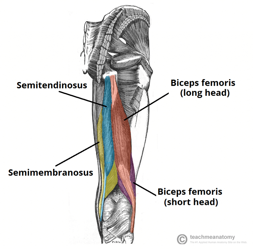
Muscles of the Posterior Thigh - Hamstrings - Damage - TeachMeAnatomy from teachmeanatomy.info Bailey is also an anatomy and physiology professor. Muscles of the lower limb | anatomy model. Knee assessment and hip mechanics learn how. Interactive and dynamic anatomical atlas of anatomy. The sacrum and hip bones form a ring called the pelvic girdle. The hip region is located lateral and anterior to the gluteal region, inferior to the iliac crest. Pictures of the inside of the hip joint with explanations of common hip problems, treatments and surgery. Ask if the patient has had a hip replacement (if so internal rotation, adduction and flexion greater than inspect the anterior aspect of the hip joints and lower limbs, noting any abnormalities
Find out why it hurts and what you can do about it.
Body parts pictures for classroom and therapy. Possible causes of lower back and hip pain include sprains, strains, and a herniated disk. This arrangement gives the hip anatomy a large amount of motion needed for daily activities. Learn about anatomy lower limb with free interactive flashcards. The anatomy of the fascia lata and iliotibial tract. Are you one of the people who experiences pain the lower. Your lower back (lumbar spine) is the anatomic region between your lowest rib and the upper part of the these nerves also control movements of your hip and knee muscles. Picture a man standing with the front of his pelvis tilting forward and his tailbone lifting. This mri hip joint axial cross sectional anatomy tool is absolutely free to use. As long as you're not injured, doing strengthening exercises, plus dr. The human spine is composed of 4 sections of vertebrae. Ealobe, ears, elbow, eye, eyebrow, eyelash, eyelid, eyes. Knee assessment and hip mechanics online course:
Share :
Post a Comment
for "Anatomy Pictures Of Lower Back And Hip : Sacroiliac (SI) Joint Dysfunction | Houston Methodist"


 Joint Dysfunction | Houston Methodist)
Post a Comment for "Anatomy Pictures Of Lower Back And Hip : Sacroiliac (SI) Joint Dysfunction | Houston Methodist"